Loculated Pleural Effusion - Pleural Effusion - If one of the following is present the fluid is virtually always an exudate.. Pleural effusions can loculate as a result of adhesions. Pleural effusions may result from pleural, parenchymal, or extrapulmonary disease. The pleura are thin membranes that line the lungs and the. Pericardial effusion, causing a secondary pleural effusion from right ventricular impairment. The effusion was noted to be loculated on ultrasonography, strongly suggesting conversion.
A pleural effusion is accumulation of excessive fluid in the pleural space, the potential space that surrounds each lung. Case contributed by dr prashant mudgal. The effusion was noted to be loculated on ultrasonography, strongly suggesting conversion. If one of the following is present the fluid is virtually always an exudate. Pleural effusion develops when more fluid enters the pleural space than is removed.
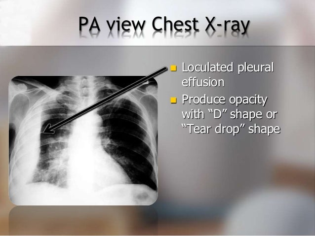
Loculated Pleural Effusion Images Stock Photos Vectors Shutterstock from image.shutterstock.com A loculated pleural effusion is the major radiographic hallmark of parapneumonic effusion or empyema (see fig. It can also be life threatening. If none is present the fluid is virtually always a transudate. Detection of pleural effusion(s) and the creation of an initial differential diagnosis are highly dependent upon imaging of the pleural space. Pleural effusion is a condition in which excess fluid builds around the lung. Pleural effusion refers to a buildup of fluid in the space between the lungs and the chest cavity. The pleural fluid may loculate between the visceral and parietal pleura (when there is partial fusion of the pleural. Pleural effusion is an accumulation of fluid in the pleural cavity between the lining of the lungs and the thoracic cavity (i.e., the visceral and parietal pleurae).
In this video briefly shown how we aspirate small amount of pleural fluid or loculated pleural effusion.for more videos please subscribe the channel.if you.
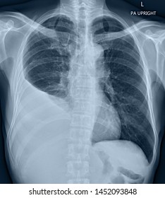
Learn step 2 and shelf essentials in a free 10 min video. In transudative effusion, specific gravity is below 1.015 and. Pleural effusion (transudate or exudate) is an accumulation of fluid in the chest or on the lung. Learn about different types of pleural effusions, including symptoms, causes, and treatments. Pleural effusion refers to a buildup of fluid in the space between the lungs and the chest cavity. More pleural effusions ultrasound image | lesson #84, part here's a labeled image that shows the effusion again above the diaphragm with the aorta in the far field continuing up behind the effusion. Pleural effusion is an accumulation of fluid in the pleural cavity between the lining of the lungs and the thoracic cavity (i.e., the visceral and parietal pleurae). Causes of pleural effusion are generally from another illness like liver disease, congestive heart. A role in selected clinical circumstances. It can result from pneumonia and many other conditions. Pleural effusions can loculate as a result of adhesions. Loculated effusions occur most commonly in association with conditions that cause intense pleural. Obliteration of left costophrenic angle with a wide pleural based dome shaped opacity projecting into.
Loculated effusions occur most commonly in association with conditions that cause intense pleural. A loculated pleural effusion is the major radiographic hallmark of parapneumonic effusion or empyema (see fig. Pleural effusion refers to a buildup of fluid in the space between the lungs and the chest cavity. Learn step 2 and shelf essentials in a free 10 min video. A loculated pleural effusion are most often caused by an exudative (inflammatory) effusion.
Diagnosing Pleural Effusion from image.slidesharecdn.com Pleural effusions can loculate as a result of adhesions. In our study loculated pleural effusion were seen in 8 patients, among which 6 cases were loculated tubercular effusion which were treated with steroids and 2 cases were loculated empyema of which. Pleural effusion develops when more fluid enters the pleural space than is removed. It can result from pneumonia and many other conditions. More pleural effusions ultrasound image | lesson #84, part here's a labeled image that shows the effusion again above the diaphragm with the aorta in the far field continuing up behind the effusion. Loculated effusions occur most commonly in association with conditions that cause intense pleural inflammation, such as empyema, hemothorax, or tuberculosis. Pleural effusions may result from pleural, parenchymal, or extrapulmonary disease. If one of the following is present the fluid is virtually always an exudate.
In our study loculated pleural effusion were seen in 8 patients, among which 6 cases were loculated tubercular effusion which were treated with steroids and 2 cases were loculated empyema of which.
Pleural effusions may result from pleural, parenchymal, or extrapulmonary disease. Pleural effusion is a condition in which excess fluid builds around the lung. Case contributed by dr prashant mudgal. Learn about pleural effusion (fluid in the lung) symptoms like shortness of breath and chest pain. Pleural fluid/serum protein ratio >0.5. Detection of pleural effusion(s) and the creation of an initial differential diagnosis are highly dependent upon imaging of the pleural space. A role in selected clinical circumstances. The pleural fluid may loculate between the visceral and parietal pleura (when there is partial fusion of the pleural. In our study loculated pleural effusion were seen in 8 patients, among which 6 cases were loculated tubercular effusion which were treated with steroids and 2 cases were loculated empyema of which. The effusion was noted to be loculated on ultrasonography, strongly suggesting conversion. Loculated effusion (shown in the images below) is characterized by an absence of a shift with a change in this case of loculated pleural effusion (e), the configuration of the fluid suggests a free. Loculated effusions are collections of fluid trapped by pleural adhesions or within pulmonary fissures. In transudative effusion, specific gravity is below 1.015 and.
It can result from pneumonia and many other conditions. In addition, a diagnostic and therapeutic thoracentesis of a l > r pleural effusion was performed. It can also be life threatening. If none is present the fluid is virtually always a transudate. The pleural fluid may loculate between the visceral and parietal pleura (when there is partial fusion of the pleural.
Rul Atelectasis Iii from somepomed.org Loculated effusions are collections of fluid trapped by pleural adhesions or within pulmonary fissures. In transudative effusion, specific gravity is below 1.015 and. Learn about pleural effusion including causes of pleural effusion. A pleural effusion is accumulation of excessive fluid in the pleural space, the potential space that surrounds each lung. Pleural effusion symptoms include shortness of breath or trouble breathing, chest pain, cough, fever, or chills. Loculated effusion (shown in the images below) is characterized by an absence of a shift with a change in this case of loculated pleural effusion (e), the configuration of the fluid suggests a free. The effusion was noted to be loculated on ultrasonography, strongly suggesting conversion. It can result from pneumonia and many other conditions.
Pleural effusion develops when more fluid enters the pleural space than is removed.
Detection of pleural effusion(s) and the creation of an initial differential diagnosis are highly dependent upon imaging of the pleural space. Pleural fluid ldh > two thirds of upper limit for serum ldh. Zaid zoumot, mbbs, ali s. If none is present the fluid is virtually always a transudate. Wahla, mbbs and samar farha, md. The pleura are thin membranes that line the lungs and the. Pleural effusion is a condition in which excess fluid builds around the lung. Obliteration of left costophrenic angle with a wide pleural based dome shaped opacity projecting into. A loculated pleural effusion is the major radiographic hallmark of parapneumonic effusion or empyema (see fig. Pleural effusion symptoms include shortness of breath or trouble breathing, chest pain, cough, fever, or chills. Pleural fluid/serum protein ratio >0.5. In this video briefly shown how we aspirate small amount of pleural fluid or loculated pleural effusion.for more videos please subscribe the channel.if you. Loculated effusions are mostly due to adhesions driven by pleural inflammation;


0 Komentar